Fig. 2.

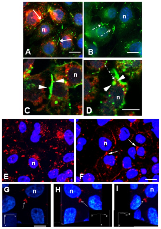
Immunocytochemical localization of Cx43 gap junction protein in control and dynasore-treated cells. (A–E) Cells were treated with 80 µM dynasore (B,D) or diluent (A,C,E) for 1 hour. Transferrin (red) uptake (A,B) was minimal in cells treated with dynasore (B) compared with that seen in controls (A). Note the colocalization (yellow) of dynamin (red in C,D) and Cx43 (green) in an area of the gap junction buds (arrowheads). Typical linear gap junction plaques, some of which had small buds, were evident in control populations (A), whereas gap junction plaques with relatively large gap junction buds (dashed arrows) were prevalent in dynasore-treated cell populations (B). (F–I) In the siRNA dynamin knockdown cell populations, there was decrease in the number of annular gap junctions and an increase in the number of gap junction buds (arrows) in cells compared with control populations (E). The attachment of a gap junction bud was demonstrated with confocal immunocytochemical 3D-reconstruction in siRNA knockdown cells (G–I). Inserts represent the rotation around the y-axis; n, nucleus. Scale bars: 7 µm (A,B); 10 µm (C,D,G–I); 20 µm (E,F).
