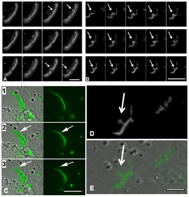
Fig. 5.
Time-lapse imaging montage of gap junction plaque endoexocytosis in cells transfected to express Cx43-GFP. (A,C) Note the relatively rapid formation and release of gap junction buds from the plaque in the control population (arrows). (B,D,E) In cells treated with dynasore, the gap junction bud (arrows) formed and then remained tethered to the gap junction plaque for the duration of the imaging period (7 hours). Corresponding fluorescence and DIC images demonstrate budding (C,E). The bud in B is enlarged in D. See supplementary material Movies 2 and 3. Scale bars: 2.50 µm (A); 5 µm (B); 3 µm (C); 2 µm (D,E).

