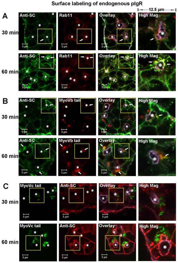
Fig. 6.
pIgR endocytosed from the basolateral membrane enters the transcytotic pathway but does not intersect with the regulated secretory pathway. (A) Non-transduced LGACs were incubated with sheep anti-rabbit SC antiserum for 1 hour at 4°C, rinsed and incubated at 37°C. Cells were fixed at the time points indicated, permeabilized, and labeled with primary mouse anti-Rab11 antibody, secondary Alexa Fluor®-488-conjugated donkey anti-sheep and Alexa Fluor®-568-conjugated goat anti-mouse antibodies, and Alexa Fluor® 647-conjugated phalloidin. LGACs expressing mCherry-myosin Vb tail (B) or EGFP-myosin Vc tail (C) were incubated with sheep anti-rabbit SC antiserum for 1 hour at 4°C, rinsed and incubated at 37°C. Cells were fixed at the time points indicated, permeabilized, and labeled with secondary Alexa Fluor®-488-conjugated donkey anti-sheep antibody (B) or Alexa Fluor®-568-conjugated donkey anti-sheep antibody (C), and Alexa Fluor®-647-conjugated phalloidin. Actin is displayed in purple in overlay and high magnification images. White arrows, colocalization between red and green; *, lumena. Scale bars: 5 µm.

