Figure 1.
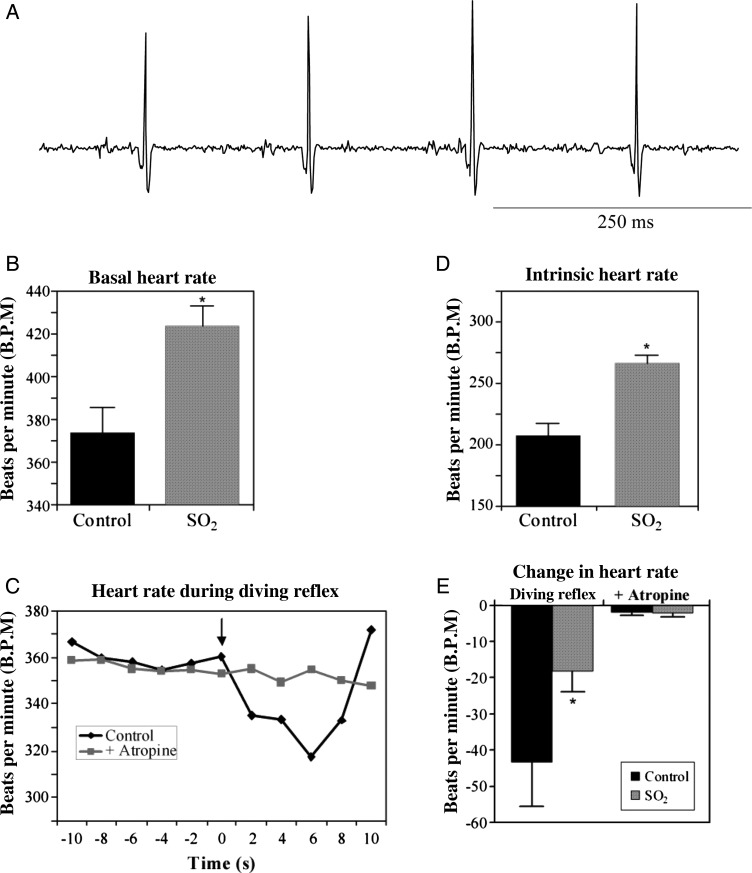
Representative ECG trace recorded from a P5 rat pup (panel A). R–R waves from ECGs were used to determine baseline heart rate (B). SO2-exposed animals exhibited a 13.4% increase in the basal heart rate compared with control pups (SO2, 424 ± 9 b.p.m.; n = 10; control, 374 ± 12 b.p.m.; n = 11; *P < 0.05). The diving reflex was used to examine the reflex control of parasympathetic activity to the heart in P5 pups (C). The basal HR was significantly decreased upon the activation of the diving reflex by placing one to two drops of cold (10°C) water on the pup's nose (indicated by the arrow; black line). Following blockade of parasympathetic activity by administering the muscarinic antagonist atropine sulfate (1 mg/kg, sc), cold water was no longer able to elicit a decrease in HR (grey line). Individual R–R intervals recorded from an example ECG were calculated, converted to HR, and averaged for every 2 s. This response was compared between control and SO2-exposed pups (E). While the diving reflex significantly decreased HR in control animals (−43 ± 6 b.p.m.; n = 11; P < 0.05), the response was significantly blunted in SO2-exposed pups (−18 ± 6 b.p.m.; n = 10; P > 0.05), and abolished by atropine sulfate. The intrinsic HR was isolated by administering atenolol (10 mg/kg, sc) after atropine sulfate (D). SO2-exposed animals exhibited a significantly increased intrinsic HR (266 ± 7 b.p.m.; n = 10) compared with control pups (208 ± 10 b.p.m.; n = 11; *P < 0.05).

