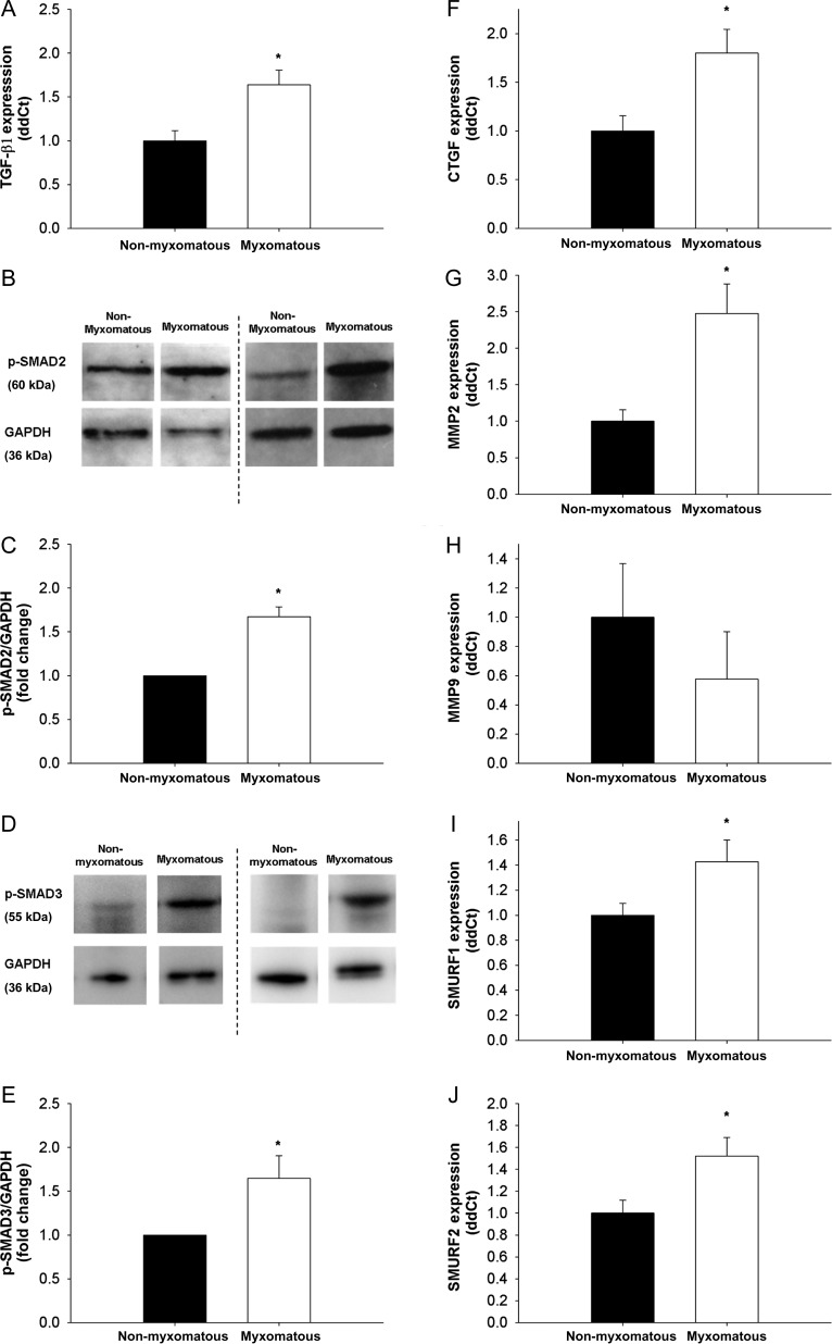
Figure 1.
TGF-β1 expression, levels of canonical TGF-β1 signalling molecules, and SMAD target gene expression in non-myxomatous and myxomatous human mitral valve tissue. (A) TGF-β1 expression is significantly increased in MMVD (qRT–PCR, n= 24 non-myxomatous valves, n= 24 myxomatous valves). (B–E) Western blots showing SMAD2/3 phosphorylation (B and D) and subsequent quantitation using densitometry (C and E). Note that SMAD2/3 phosphorylation—indicative of canonical TGF-β1 signalling—is significantly increased in MMVD (n= 5 non-myxomatous valves, n= 5 myxomatous valves). (F–H) Changes in CTGF, MMP2, and MMP9 in MMVD. Note that CTGF (F) and MMP2 (G) are markedly increased in MMVD (qRT–PCR, n= 24 non-myxomatous valves, n= 24 myxomatous valves). (I and J) Changes in expression of the intracellular E3 ubiquitin ligases SMURF1 and SMURF2, which are key negative regulators of canonical Smad signalling. Note that both SMURF1 and SMURF2 are markedly increased in human MMVD (n= 24 non-myxomatous valves, n= 24 myxomatous valves). *P < 0.05 in all figures.

