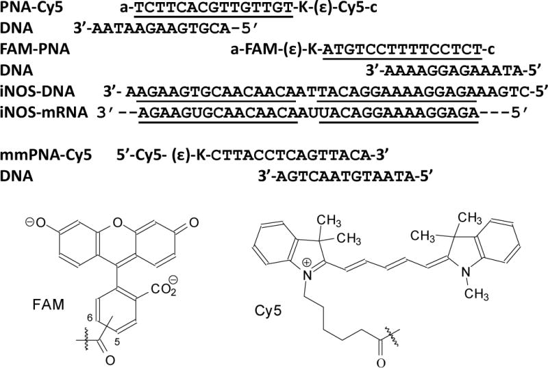
Fig. 2.

PNA•DNA FRET probes used in this study. The sequences of the PNAs are shown with their orientation indicated by “a” for the amino terminus and “c” for the carboxy terminus, and the DNAs with the 5’- and 3’-ends as indicated. The sections of complementarity are underlined. The donor and acceptor fluorophores shown below are connected through the indicated carboxy group to the ɛ-amino terminus of the lysines on the PNA.
