
Figure 3A.
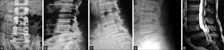
Degenerative instability managed by PLIF; (a) X-Ray lumbosacral (L/S) spine Anteroposterior view. (b) X-Ray L/S spine lateral view showing degenerative spondylolisthesis at L4 over L5. (c) Dynamic film in flexion showing anterior translation and angular displacement. (d) Dynamic film in extension showing differences in translation and angular motion. (e) T2W MRI scan of that patient with degeneration and spondylolisthesis at L4/5 level.