
Figure 4B.
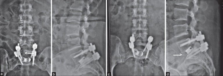
(a) Postoperative X-ray lumbosacral spine anteroposterior view and (b) lateral view showing good implant position and the transforaminal approach (arrow). (c) 1 year followup X-Ray anteroposterior view and (d) lateral view showing listhesis reduction and radiological fusion (arrow)