
Figure 1.
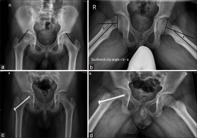
X-ray pelvis with both hip joints (a) Anteroposterior view of a 15-year-old boy showing a mild slip. The blurring of physis (Blanch sign) and capital epiphysis dipping below the Klein's line are observed. (b) Frog leg lateral view of the same child showing slip angle (Southwick) measuring 20° on the right side. (c and d) AP and frog leg lateral views showing a centrally placed cannulated screw crossing the physis