
Figure 1.
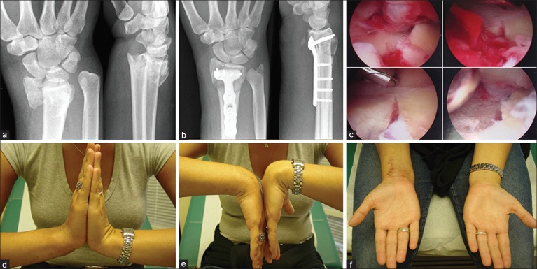
(a) Radiographs showing preoperative wrist posteroanterior and lateral views of patient 1 showing an intraarticular C2 type distal radial fracture, (b) Postoperative radiograph of wrist showing posteroanterior and lateral views of the same patient, following stable fixation of distal radial fracture with a volar fixed angle locking plate, (c) Photograph showing wrist arthroscopic views. A shaver is being used for clearing the hematoma and debris in the joint, while the articular reduction is confirmed, (d-f) Clinical photographs showing range of motion at the final followup