Fig. 3.
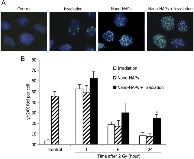
The effect of nano-HAPs on the γH2AX foci caused by irradiation. U251 cells growing in chamber slides were exposed to 10 mg/L nano-HAPs for 1 h, irradiated (2 Gy), fed fresh medium, and fixed at the specified times for immunocytochemical analysis for nuclear γH2AX foci. Foci were determined in 50 nuclei per treatment per experiment. (A) Representative micrograph images gained from the control cells and cells that had received 10 mg/L nano-HAPs alone for 1 h, 2 Gy irradiation alone, and nano-HAPs (10 mg/L; 1 hr before irradiation) + irradiation at 24 h after irradiation (2 Gy). (B) Vehicle-treated cells (empty columns), cells treated with nano-HAPs alone (hatched columns), and cells treated with the combination of nano-HAPs + irradiation (2 Gy; filled columns). Cells with >5 foci per nucleus were classified as positive for radiation-induced γH2AX. *P < .01 according to Student's t-test (irradiation vs nano-HAPs + irradiation).

