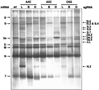
FIG. 3.
Northern blot analysis of rTGEVs. ST cells were infected with rTGEV at an MOI of 0.5 (for the wild type [wt] and CS-B mutants) or 1 (for CS-L and double mutants). Total RNA was extracted at 20 hpi and analyzed by Northern blotting with a probe complementary to the 3′ end of the gRNA. To normalize the amount of viral RNA in the gel, lanes L and D were loaded with three times the amount of the other lanes. L, CS-L mutant; B, CS-B mutant; D, double mutant. Viral mRNAs are indicated on the left side of the figure, and new sgmRNAs that have been clearly identified are indicated on the right (some of them correspond to the alternative sgRNAs analyzed in this work, indicated by the same number). n.i., still unidentified sgmRNAs.

