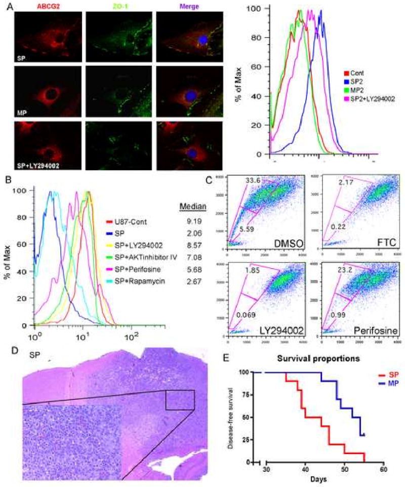
Figure 5. PI3K/Akt pathway regulates ABCG2 activity in U87MG stable cell line and the SP phenotype of human neurospheres.

(A) Immunostaining for hABCG2 (red) shows a membrane staining in SP2 cells versus a cytoplasmic staining in MP2 cells; ZO-1 (green) delimitate the membrane. In the right panel, FACS analysis using cell surface hABCG2 antibody confirms high staining intensity in SP2 cells. Blocking of PI3K with LY294002 induced a change in ABCG2 localization, from the cell membrane to the cytoplasm.
(B) Mitoxantrone exclusion assay in SP2 cells after incubation with LY294002, Akt inhibitor IV, perifosine and rapamycin. Blocking of the PI3K and Akt pathways abolished the ability of ABCG2 to efflux mitoxantrone.
(C) Hoechst staining of human neurosphere culture shows the presence of a SP that was blocked by FTC. Treatment with LY29004 totally abolished the SP fraction while perifosine displayed a stronger effect in blocking the lower SP fraction.
(D) H&E of nude mice injected with SP cells isolated from human GBM neuropsheres.
(E). Kaplan-Meier survival curve shows that SP cells isolated from human neurospheres are more tumorigenic then MP cells.
