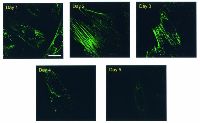
FIG. 1.
Staining of cellular actin cytoskeleton in live EIAV-infected ED cells by fluorescent phallacidin. EIAVuk-infected ED cells grown on Lab-Tek chambers were incubated at 37°C for 20 h with 25 μM BODIPY FL phallacidin. The fluorescent drugs were then replaced with drug-free culture medium. Images of fluorescent actin filaments were captured daily over a 5-day period by using Leica confocal microscope and identical imaging conditions. Bar, 15 μm.

