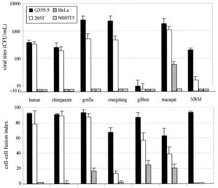
FIG. 2.
Infection and cell-cell fusion activities and host range of the HERV-FRD and simian orthologous envelopes. Upper panel: infection of feline (G355-5), human (293T and HeLa), and murine (3T3) cells by SIV-based particles pseudotyped with the indicated ERV-FRD envelopes. Experimental conditions were the same as in Fig. 1, with negative and positive control values as in Table 1. NWM, New World monkey (Callithrix jacchus marmoset). Lower panel: cell-cell fusion assay for the ERV-FRD envelopes and cells as in the upper panel. The G355-5 and HeLa cells were transfected using Lipofectamine (Invitrogen; 2 μg of DNA for 5 × 10⁵ cells), and the 293T and TE671 cells were transfected using calcium phosphate precipitation (Invitrogen; 5 μg of DNA for 5 × 10⁵ cells). Fusion activities were measured 12 to 36 h posttransfection of the corresponding expression vectors. To visualize syncytia, cells were fixed in methanol and stained by adding May-Grünwald and Giemsa solutions (Sigma) according to the manufacturer's instructions. The fusion index, which represents the percentage of fusion events in a cell population, is defined as [(N − S)/T] × 100, where N is the number of nuclei in the syncytia, S is the number of syncytia, and T is the total number of nuclei counted.

