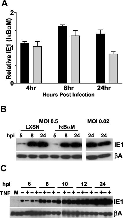
FIG. 7.
Inhibition of the canonical NF-κB pathway does not inhibit expression of HCMV IE1. (A) NHDF-LXSN or -IκBαM cells were infected at an MOI of 0.005 (dark bars) or 0.1 (light bars) and were harvested at various hours postinfection (hpi) for analysis of ie1 expression by real-time PCR. Results represent the relative amount of ie1 expression in IκBαM cells compared to that of control cells (LXSN) at the various times. Values are averaged data from three independent experiments, and error bars represent the standard errors of the means. (B) NHDF-LXSN or -IκBαM cells were infected with HCMV at an MOI of 0.5 or 0.02. Cells were harvested at various hpi for analysis of IE1 protein levels by Western blotting. For samples treated at an MOI of 0.02, LXSN-infected cells are shown in the left lane, and IκBαM cells are shown in the right. (C) NHDF-LXSN (first two lanes of each time point) or -IκBαM cells (second two lanes of each time point) were infected with HCMV at an MOI of 2, and cells were harvested at various hpi for analysis of IE1 protein levels by Western blot. For each time point, cells were either treated with (+) 1 nM TNF or were not (−).

