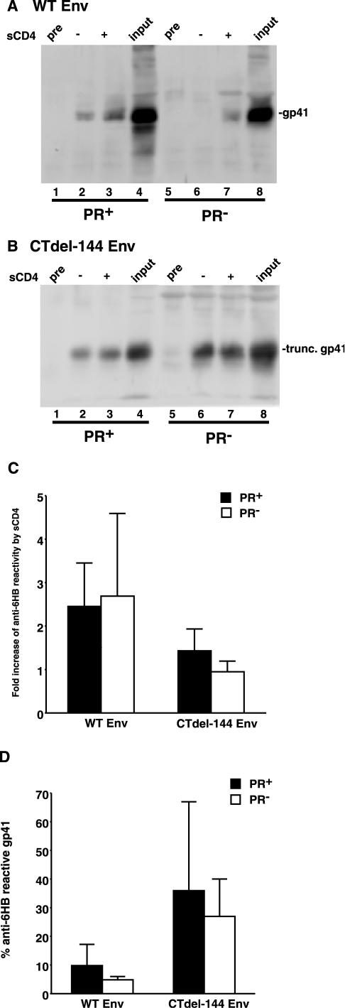
FIG.4.
PR+ and PR− virions display comparable levels of 6HB formation. Concentrated HIV-1 virions (prepared as described in the legend of Fig. 1, except that in this case NL4-3 [1] and CTdel-144 [39] and their PR− counterparts were used) were incubated with an anti-6HB antiserum (rabbit serum no. 948) or preimmune serum (pre) in the presence or absence of 1 μg of sCD4 (Immuno Diagnostics) at 37°C for 2 h. Virions were then pelleted by centrifugation (20,000 × g for 2 h) prior to lysis and immunoprecipitation with protein G Sepharose beads. The immunoprecipitated material was separated by sodium dodecyl sulfate-polyacrylamide gel electrophoresis and subjected to quantitative Western blotting with the anti-gp41 MAb T32. An amount of viral lysate equivalent to input virus was run as a positive control. Gels were run for WT Env (A) and CTdel-144 Env (B). The increase in anti-6HB reactivity (n-fold) induced by sCD4 in the PR+ (solid bar) and PR− (open bar) context (C) is shown. Also shown is the percentage of anti-6HB-reactive gp41, calculated by dividing the amount of 6HB-reactive gp41 following sCD4 addition by input gp41 in PR+ (solid bar) and PR− (open bar) virions (D). Rabbit serum no. 948 was obtained from C. Weiss (12, 23). Data are means ± standard deviations of the results of at least three independent experiments.

