
Figure 1.
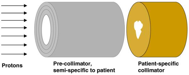
Geometry for the simplified Monte Carlo simulations. A uniform mono-energetic 235 MeV proton beam is incident on the face of the pre-collimator, which is upstream of the patient-specific collimator which is, in turn, immediately upstream of a cylindrical homogeneous tissue-equivalent phantom (to the right, not shown). The pre-collimator prevents unnecessary protons from impinging on the patient-specific collimator, while the final collimator provides the patient-specific collimation, but also shields the patient from neutrons produced in the pre-collimator. For these calculations, both the collimator and pre-collimator had circular internal and external diameters, respectively, of 50 and 113 mm. Doses due to neutrons originating in the collimator were tallied in 10 mm thick slices along the length of a cylindrical homogeneous tissue-equivalent phantom (1.5 m length × 0.5 m diameter), axis perpendicular to the proton-beam direction, located immediately downstream of the patient-specific collimator.