
Figure 2.
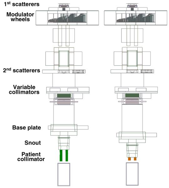
Simulations of the proton therapy treatment heads at the gantry beamlines at MGH, with a 65-mm thick brass collimator (above), and an optimized plastic or metal/plastic collimator (below). The proton beam is incident from the left, and the modulated collimated beam is incident on a water phantom on the right. Beam and treatment-head settings were chosen to represent a typical field, with a prescribed range of 157 mm, a modulation width of 68.5 mm, and an aperture opening diameter of 60 mm. The area irradiated with protons at the base plate position corresponds to a diameter of roughly 250 mm, depending on the setting of variable collimators upstream of the snout retraction area. The treatment head components are described in detail elsewhere (Paganetti et al 2004).