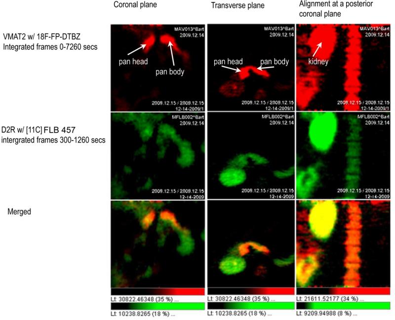
Figure 3.
Papio Ursinus was imaged twice in one session for a study comparing the region of interest defined by a VMAT2 probe, 18F-FP-(+)-DTBZ, and a D2R probe [11C] FLB 457.The [11C] study was performed first and activity from the probe allowed to decay for six half lives (11C T1/2 = 21 minutes) before injection of the secondVMAT2 probe, 18F-FP-(+)-DTBZ. Following reconstruction of the images the pancreatic region of interest was defined using the VMAT2 probe. The overlap of the D2R and VMAT2 signals was then examined (bottom row-Merged). All studies were approved by the Columbia Institutional Animal Care and Use Committees.

