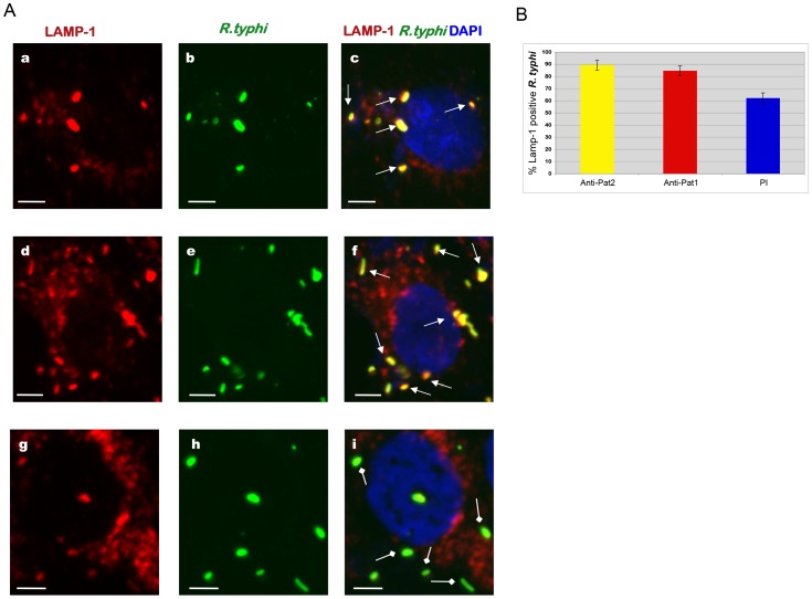
Figure 9. Effect of anti-Pat1 and anti-Pat2 pretreatment on R. typhi phagosome escape by IFA.
Rickettsiae were incubated with 2 µg of affinity purified anti-Pat1, anti-Pat2 or pre-immune IgG (PI) for 30 min on ice. Pretreated rickettsiae were added onto Vero76 monolayer followed by incubation at 34°C and 5% CO2. At 30 min postinfection, the cells were fixed with 4% paraformaldehyde and immunolabeled with anti-LAMP-1 (at 1∶100 dilution) mouse monoclonal antibodies (Abcam Inc) and anti-R. typhi rat serum (at 1∶500 dilution) as primary antibodies. The anti-rat-Alexa Fluor-488 (green) and anti-mouse-Alexa Fluor-594 (red) were used as secondary antibodies. The cell nuclei were stained with DAPI (blue). Samples were viewed under a LSM5DUO confocal microscope and images were processed using ZEN imaging and analysis software. (A) Representative micrograph of anti-Pat1 (panels a to c), anti-Pat2 (panels d to f) or pre-immune IgG (panels g to i) treated rickettsiae that were infected of Vero76 cells. The white arrows show rickettsiae in LAMP-1 positive phagosome, with the square head arrow showing rickettsiae escaping or escaped from the phagosome. Scale Bar = 5 µm. (B) Quantitation of percent rickettsiae in LAMP-1 positive phagosome was determined as percent LAMP-1 positive R. typhi by scoring 100 bacteria for each treatment. Each treatment was repeated four times. Error bars represent standard error of the means. Percent LAMP-1 positive R.typhi for anti-Pat1 or anti-Pat2 pretreatment was found to be significantly different (P<0.05[two-tail t-test]) from that of PI.

