
Figure 9. In situ zymography of midgut gland.
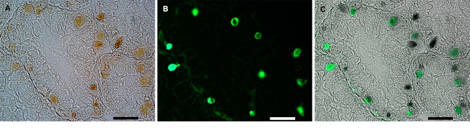
A. Light microscopy of several tubulo-acini, showing numerous pigmented endosymbiotic corpuscles. B. Green fluorescence (FITC) indicates substrate degradation (DQ-gelatin). C. Merging of A and B showing substrate degradation by most endosymbiotic corpuscles. Corpuscles which do not show fluorescence may be either dead symbionts or, more likely, they may be the result of differences in the penetration of reagents. Scale bars = 50 µm.