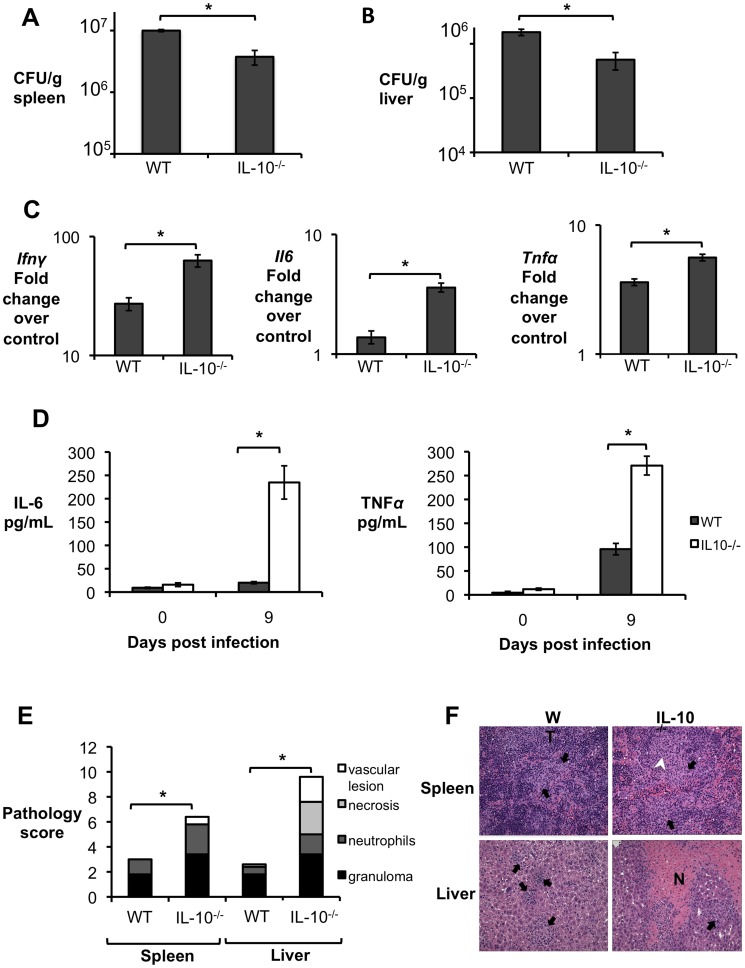
Figure 2. Lack of IL-10 results in lower bacterial survival and increased pathological changes during early Brucella abortus infection in vivo.
(A,B) Colonization of spleen (A) and liver (B) of control or IL-10−/− mice by B. abortus at 9 d.p.i. (C) qRT-PCR analysis of IFN-γ, IL-6 and TNF-α gene expression in spleens of C57BL/6 and IL-10−/− mice infected with B. abortus 2308 for 9 days (similar results for liver). (D) IL-6 and TNF-α levels measured by ELISA in serum of C57BL/6 and IL-10−/− mice infected with B. abortus 2308 for 9 days. n = 5. Values represent mean ± SEM. *P<0.05 using unpaired t-test statistical analysis.( E) Histopathology score of spleen and liver from C57BL/6 and IL-10−/− mice infected with B. abortus 2308 for 9 days. (F) Representative pictures from (E). Black arrow shows microgranulomas, white arrowheads shows neutrophilic infiltrate and upper case N shows areas of coagulative necrosis (×20). n = 5. Values represent individual mice (black circles) and geometric mean (black dash). *P<0.05 using Mann-Whitney statistical analysis.

