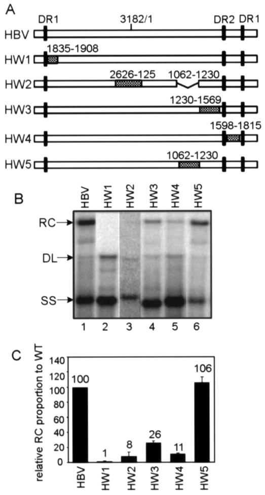
FIG. 4.
Replication of HBV/WHV chimera variants in HepG2 cells. (A) WT HBV and HBV/WHV chimera variants (HW1 to HW5). The white boxes represent HBV sequences, and the hatched boxes represent the substituted WHV sequences. The black vertical bars indicate the positions of DR1 and DR2 sequences. In the HW2 construct, HBV sequences from nt 1062 to 1230 were deleted to maintain the genome size. (B) Southern blotting of WT HBV and HBV/WHV chimeras. Replicative intermediates were isolated from the cytoplasmic viral capsids in HepG2 cells and analyzed by Southern blotting. A genome-length, minus-strand-specific RNA probe was used to detect viral DNA. The positions of RC DNA, DL DNA, and SS DNA are indicated. (C) Proportions of RC DNA of the chimeras relative to that of the WT standard. The proportion of RC DNA is the level of RC DNA among the total intermediates containing mature minus-strand DNA on Southern blots. In each transfection, the RC DNA proportion of each of the chimeras was normalized to that of the WT standard, which gives the value for the relative RC DNA proportion. The mean values of the relative RC DNA proportion from at least six independent transfections were plotted on the histogram. The numbers above each bar indicate the mean values of the relative RC proportion. The error bars indicate the standard deviations.

