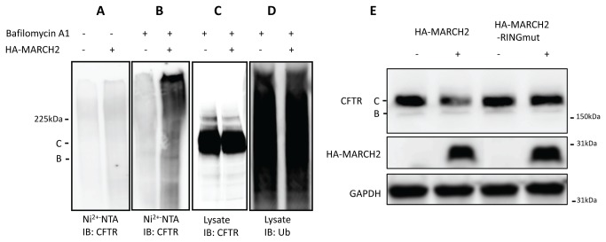
Figure 4. In vivo ubiquitination of CFTR by MARCH2.
(A) HEK293 cells were first co-transfected with 3µg CFTR (pCMV-CFTR) and 3µg His6-ubiquitin. After 24 h, the cells were transfected again with 1µg HA-MARCH2 or the vector plasmid pRK5KS. Twenty-four hours after the second transfection, cell lysates were harvested under denaturing conditions (in a buffer containing 6 M guanidinium hydrochloride). Ubiquitinated proteins were affinity-purified with Ni2+-NTA-agarose beads, eluted with Laemmli buffer, separated on SDS-PAGE, transferred to a PVDF membrane, and subjected to the immunoblot analysis with anti-CFTR mouse monoclonal antibody. (B) Same as in (A) except cells were treated with 400nM bafilomycin to inhibit lysosome activity 24 hours before harvest under denaturing conditions. Ubiquitinated proteins were affinity-purified with Ni2+-NTA-agarose beads and subjected to the immunoblot analysis with anti-CFTR mouse monoclonal antibody. (C) Same as in (B) total cell lysates were subjected to immunoblot analysis with anti-CFTR antibody (D) The PVDF membrane in (C) was stripped and blotted with an anti-ubiquitinated proteins antibody FK2. Data shown are representative of at least three independent experiments. (E) HEK293 cells were co-transfected with 3µg CFTR (pCMV-CFTR) and 1µg HA-MARCH2 or HA-MARCH2 RING. Forty-eight hours after transfection, cell lysates were harvested and subjected to Western blot analysis with anti-CFTR mouse monoclonal antibody 217. Data shown are representative of at least three independent experiments.

