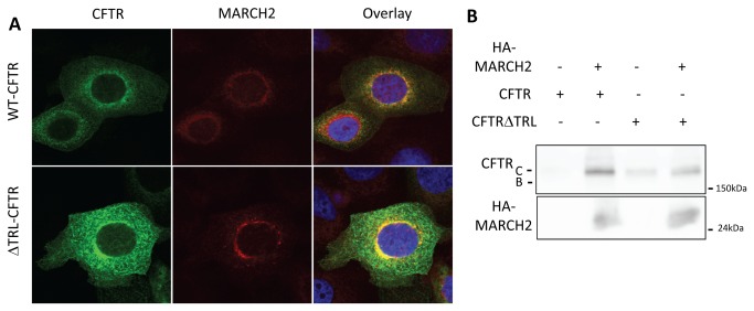
Figure 7. Localization and interaction of MARCH2 with wild-type and ΔTRL CFTR.
(A) CBFE cells grown on coverslips were co-transfected with 3µg GFP-CFTR or 3µg GFP-ΔTRL-CFTR and 0.5 µg HA-MARCH2. After 24h, the cells were fixed and subjected to indirect fluorescent immunocytochemical staining with an anti-HA mouse monoclonal antibody and anti-GFP rabbit polyclonal antibody. HA-MARCH2 appears red and GFP-CFTR green. (B) HEK293 cells were co-transfected with 3 µg GFP-CFTR or 3µg GFP-ΔTRL-CFTR and 1 µg HA-MARCH2, as indicated. After 48 h, cell lysates were harvested and immunoprecipitated with an anti-HA affinity matrix. Cell lysates and immunoprecipitated materials were subjected to immunoblot analysis with the indicated antibodies. Data shown are representative of at least three independent experiments.

