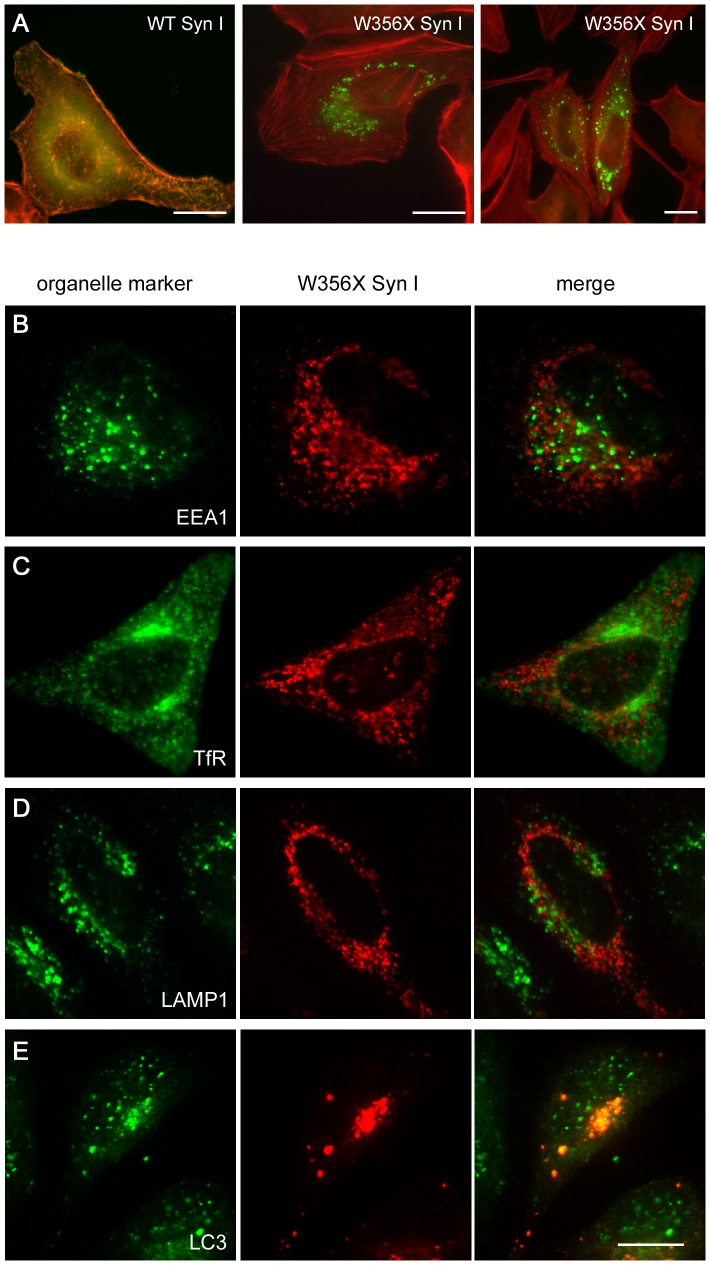
Figure 3. The W356× Syn I variant accumulates in aggregates that are labelled by an autophagosome marker.
HeLa cells were transfected with FLAG-tagged WT or W356× Syn I coding plasmids. A.Representative immunofluorescence images show that the distribution of the WT protein (green) overlaps with the distribution of the F-actin filaments labelled with phalloidin (red), while the mutant form accumulates in perinuclear aggregates. WT Syn I is expressed in the majority of the cells (not shown), while W356× Syn I is only expressed by a small fraction of the cDNA-transfected cells. B-E.W356× Syn I aggregates (red) do not co-localize with organelle markers (green) specific for either early endosomes (EEA1; B), recycling endosomes (TfR; C) or lysosomes (LAMP1; D), while they co-localize with an autophagosome marker (LC3; E). Scale bars: 15 µm.

