
Figure 4. W356× Syn I aggregates are Triton X-100 insoluble and are not ubiquitinated.
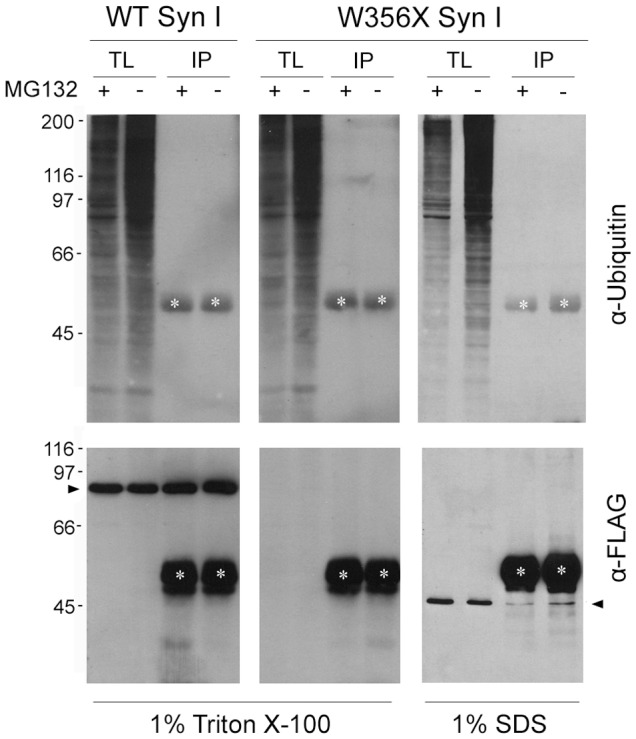
HeLa cells transfected with FLAG-tagged WT or W356× Syn I were lysed in 1% Triton X-100 or 1% SDS buffers after overnight treatment in the presence (+) or absence (−) of the proteasome inhibitor MG132 (1 µM). Total lysates (TL) were subjected to immunoprecipitation (IP) with an anti-FLAG antibody and the IP samples were analyzed by Western blotting with anti-Ubiquitin antibody to reveal protein ubiquitination (upper panels). Neither WT nor W356× Syn I appear ubiquitinated. To check for recovery of the transfected proteins after IP, the same membranes were stained with anti-FLAG antibody (lower panels). White asterisks indicate the IgG heavy chains. Black arrowheads indicate either the WT or W356× Syn I band.