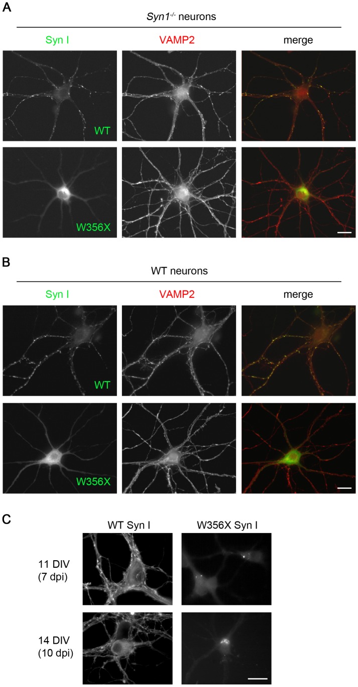
Figure 5. The W356× Syn I variant is not targeted to nerve terminals in either Syn1−/− or WT hippocampal neurons.
A. Syn1−/− hippocampal neurons were transduced at 4 DIV with lentiviruses expressing EYFP-labeled either WT or W356× Syn I (green), and fixed at 8 DIV. VAMP2 (red) was used as a marker of presynaptic contacts. B. A similar experiment was performed in WT hippocampal neurons. In both neuronal backgrounds, WT Syn I displays a punctuate pattern overlapping with VAMP2 staining, while W356× Syn I is dispersed in the cytoplasm. The presence of endogenous Syn I in WT neurons is not sufficient to correct the mis-localisation and to redirect the mutant protein to presynaptic terminals. C. Syn1−/− hippocampal neurons were transduced at 4 DIV and fixed at either 11 or 14 DIV. The prolonged expression of W356× Syn I results in the formation of somatic microaggregates. Scale bars: 20 µm.

