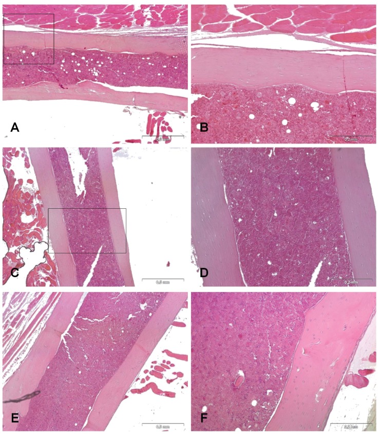
Figure 6. Histology of the femur in groups I and III and in group II.
H&E staining of longitudinal sections of femurs. Note the absence of inflammatory cells in the medullary canal, bone resorption and periosteal reaction in group I (A, B), group III (C, D), and group II (E, F) 28 days after surgery. Left panel magnification 4X (scale bars 0.5 mm); right panel magnification 10X (scale bars 0.2 mm).

