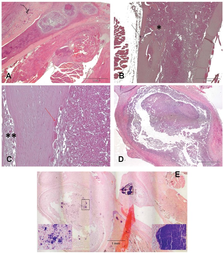
Figure 7. Histology of the femur and lymph node in group IV.
H&E staining showing inflammatory cells and micro abscesses in the medullary canal (A, 2X, scale bar 1 mm); sequestrum formation (*) and fistula (black arrow) in the cortical bone (B, 4X, scale bar 0.5 mm); endosteal bone resorption, several osteoclasts (red arrow), and marked periosteal reaction (**) (C, 20X, scale bar 0.1 mm). A lymph node reaction is shown in image D (2X, scale bar 1 mm). Image E (2X, scale bar 1 mm) showing Gram-positive staining of bacterial clusters within a macroabscess of the knee joint capsule (left small box, 40X, scale bar 0.05 mm) and within a microabscess in the medullary canal (right small box, 40X, scale bar 0.05 mm).

