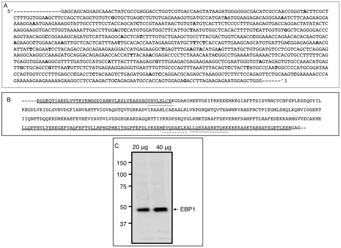
Figure 1. Nucleic acid and amino acid sequence of hamster EBP1.
(A) Partial nucleic acid sequence of the hamster EBP1 cDNA. Hamster EBP1 was around 95% similar to that of the other species. Bold face letters indicates nucleotide differences with that of the mouse EBP1. (B) Deduced amino acid sequence of the hamster EBP1 highlighting nucleolar localization signal (underline), RNA binding motifs (overline) and a transcriptional co-activator/repressor binding site (dashed overline). (C) A Western blot validating the specificity of the polyclonal EBP1 antibody. The antibody detected one approximately 48 kDa protein band in two different amounts of hamster ovarian total protein. The increase in chemiluminescence signal with higher amounts of ovarian protein indicated non-saturation of the antibody used in the analysis.

