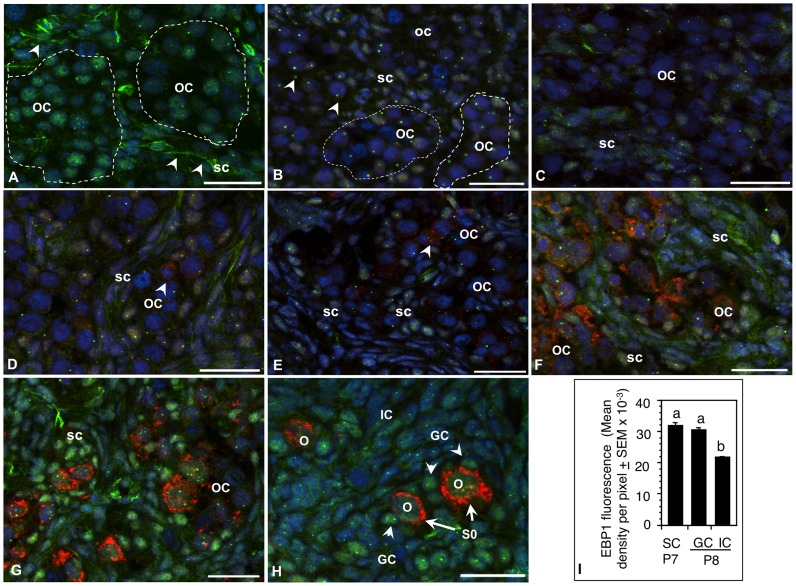
Figure 2. Spatiotemporal expression of EBP1 protein during perinatal ovary development in the hamster.
EBP1 (green) was localized in germ cells as well as undifferentiated somatic cells in the fetal ovary; however, the cell-type specific location and the degree of expression shifted to somatic cells as the ovary developed. Sections of (A) an E15, (B) a P1, (C) P3, (D) P4, (E) P5, (F) P6, (G) P7, and (H) P8 ovaries. MVH expression (red) was detectable in the oocytes in P4 ovaries (D, arrowhead) and increased appreciably with the growth of the oocytes from P6 (F) through P8 (H). Nuclei were stained with DAPI. Germ cell nests or oocyte clusters (OC) were demarcated by broken line. SC, somatic cells, O, oocytes, S0, primordial follicles, scale bar = 10 µm. (I) Quantitative representation of EBP1 immunosignal in undifferentiated somatic cells of P7 ovaries and in the granulosa cells (GC) of primordial follicles and interstitial cells (IC) of P8 ovaries. Bars with different letter = P<0.05.

