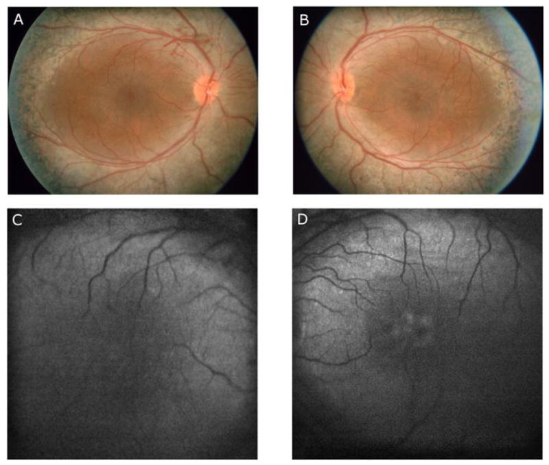
Fig. 1.

Fundus photographs obtained at the time of presentation of the right eye (A) and left eye (B), demonstrating blotchy pigment clumping along the vascular arcades and the mid-peripheral retina as well as areas of diffuse hypopigmentation for 360 degrees in the mid-peripheral retina in each eye. The left eye (B) showed cystic-appearing lesions within the macula. Autofluorescence (AF) images of the right eye (C) and left eye (D) show a ring of enhanced autofluorescence in the posterior pole in each eye. The image of the left eye (D) shows a faint ring of enhanced autofluorscece within the fovea.
