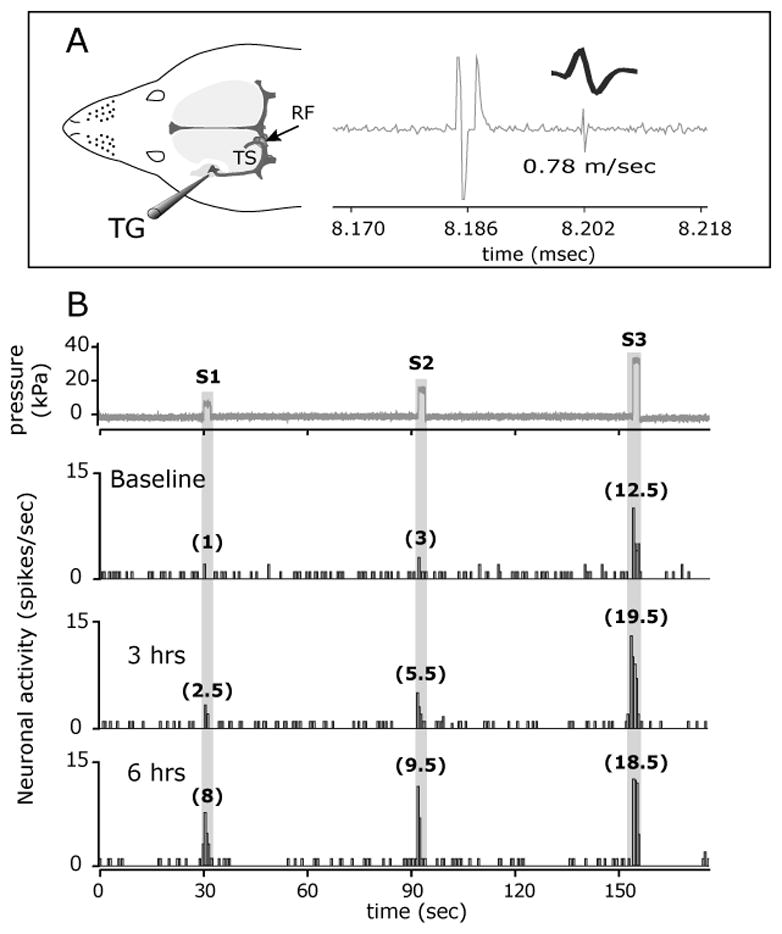
FIGURE 1.

NTG infusion evokes a delayed mechanosensitization of a C-unit meningeal nociceptor. (A) Experimental setup (left) and the identification and isolation of a C-unit meningeal nociceptor based on the response latency (16 milliseconds) to electrical stimulation of the dura and a consistent waveform (right). (B) Peri-stimulus time histograms depicting the responses to threshold (S1) and suprathreshold (S2-3) mechanical stimulation of the dura at baseline, before NTG infusion, and at 3 and 6 hrs post infusion. The numbers in parenthesis indicate mean responses to mechanical stimuli in spikes/sec. Note the progressive increase in threshold (S1) responsiveness and the lack of increases in ongoing discharge (between trials) in response to NTG. RF, receptive field; TS, Transverse sinus; TG, Trigeminal ganglion.
