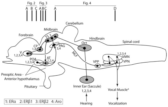
Fig. 1.
Schematic sagittal view of the plainfin midshipman (Porichthys notatus) brain showing subset of vocal (open) and/or auditory (shaded) nuclei (adapted from Goodson and Bass, 2002; Bass and McKibben, 2003; Kittelberger et al., 2006; Kittelberger and Bass, 2012). Partial shading indicates involvement in both the ascending auditory and descending vocal systems. Dots and lines represent somata and axonal projections, respectively. Connected dots and arrows represent reciprocal and unidirectional connections, respectively. Vertical bars at the top indicate approximate regions of atlas sections in Figures 2–4. Nuclei that express ERα, ERβ1, ERβ2 and aromatase are labeled 1 through 4 (see key), while those without such labels do not express either ER or aromatase. *Expression data for vocal muscle is not available. Abbreviations: AT, anterior tuberal nucleus; CP, central posterior nucleus of dorsal thalamus; MLF, medial longitudinal fasciculus; Oct, hindbrain octaval (auditory nuclei); PAG, midbrain periaqueductal gray; POA, preoptic area; TS, midbrain torus semicircularis; V, area ventralis of the telencephalon; VMN, vocal motor nucleus; VPN, vocal pacemaker nucleus; VPP, vocal prepacemaker nucleus.

