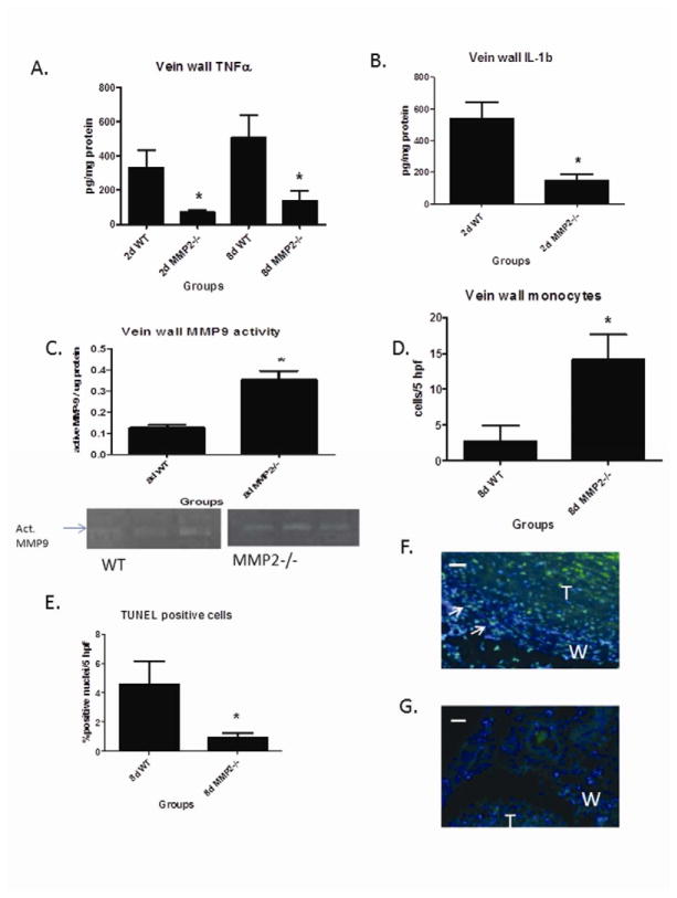
Figure 3.
Vein wall TNFα in MMP2−/− was less at 2 and 8d (A), while IL1β was reduced at 2d (B). C) Vein wall active MMP9 activity was increased in MMP2 −/− mice at 8d by zymography. D) Vein wall monocytes were increased at 8d in MMP2 −/− mice as compared to WT. E, F, G) Apoptosis as marked by TUNEL + green colocalized with DAPI blue staining nuclei showed less medial (+) cells at 8d in the MMP2 −/− vein wall compared with WT. @400X. W = wall; T = thrombus; arrows mark (+) cells; * p < .05. White measurement bar = 10 μm.

