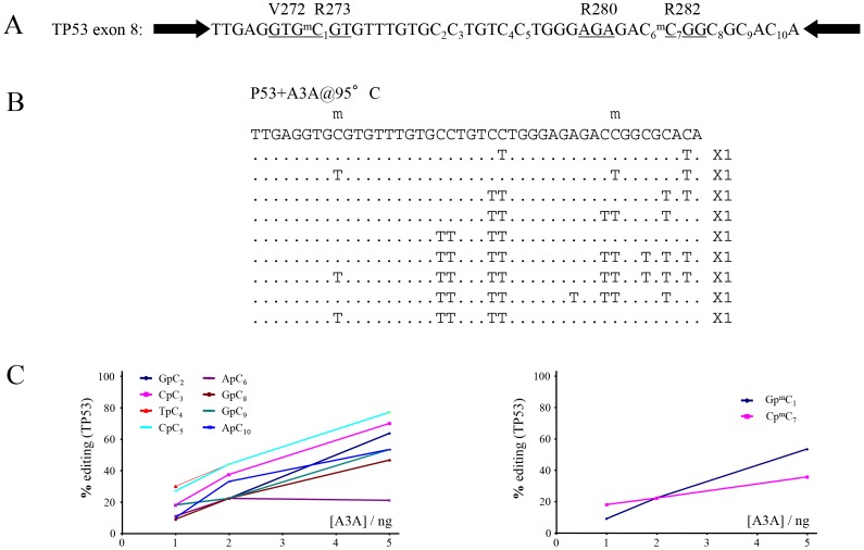
Figure 3. A3A deamination of an oligonuclotide harbouring two methylated CpGs that are TP53 mutation hotspots in cancers.
A) The TP53 target sequence is nested between two PCR primer targets (black arrows). The underlined triplets highlight the codons which are frequently substituted in cancers. B) A selection of deaminated TP53 sequences with the number of mutant sequences shown to the right. C) Site specific cytidine and 5-methyl cytidine deamination frequencies as a function of A3A concentration.

