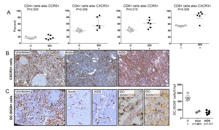
FIGURE 2.
Flow cytometric and IHC measurement of cell types in HiLNs from SIV-infected and uninfected macaques. A: Flow cytometry was performed on available samples of viably cryopreserved HiLN cells using fluorophore-conjugated antibodies to CD3, CD20, CD4, CD8, CCR5 and/or CXCR3. Shown are the percentages of CD3+/CD20− CD4+ or CD8+ lymphocytes that were also either CCR5+ or CXCR3+ from individual animals, with the group medians and interquartile ranges. Groups were compared using the Mann-Whitney nonparametric test. B: Immunohistochemistry (IHC) was performed to detect CXCR3+ cells in HiLN tissue sections, with antigen-expressing cells staining brown. Original magnification, X100 and X200 (C). C: IHC was performed to detect DC-SIGN+ cells in HiLN tissue sections (original magnifications x200). In addition, ISH for DC-SIGN mRNA was performed simultaneously on HiLN sections stained for either CD68 or MHC-II (original magnificaitons×400). The average density of DC-SIGN+ cells per 0.3mm2 was determined for individual HiLNs from the indicated disease states, with the median and interquartile ranges denoted (to the right). Group comparisons were made using the Mann-Whitney nonparametric test.

