
Figure 1. Confirmation of specificity of TLR4 knockout (TLR4-/-).
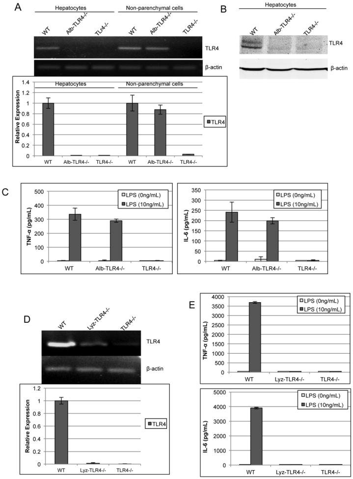
(A) RT-PCR was used to determine mRNA levels of TLR4 within either isolated hepatocytes or non-parenchymal cells (NPCs) from WT, Alb-TLR4-/-, and TLR4-/- mice. β-actin was used as endogenous control to determine relative expression compared against WT. (B) TLR4 protein levels were assessed by Western blot analysis. (C) NPCs were isolated and stimulated with LPS for 6h and the supernatant was analyzed by ELISA for either TNF-α or IL-6. (D) RT-PCR analysis of peritoneal macrophages to determine TLR4 mRNA expression in Lyz-TLR4-/-. (E) LPS response was determined in peritoneal macrophages from WT, Lyz-TLR4-/-, and TLR4-/- mice. Data represent mean ± SD. N=2 samples per group run in duplicate. Figure is representative of two experiments with similar results.