
Figure 4. Western blotting analysis using (A) combination of native electrophoresis and western blotting showing single band of cytosolic ARG I purified from the liver of H. fossilis.
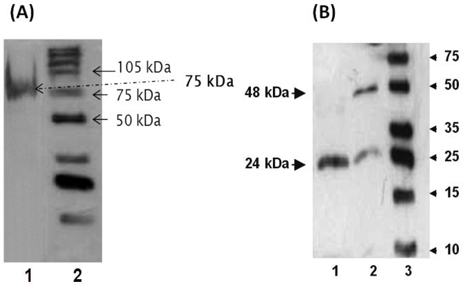
Lanes-1, Purified hepatic ARG I; 2, Molecular mass marker. (B) Western blotting showing monomer and dimers of H. fossilis ARG I. Lanes-1, sample heated at 100°C for 60 mins in sample buffer (with 200 mM DTT); 2, sample heated at 100°C for 10 min in sample buffer; 3, molecular mass marker.