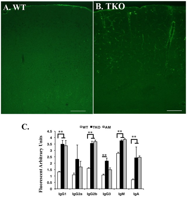
Figure 1. Autoantibodies are deposited on the microvessels of the TKO brain.
Both WT (A) and TKO (B) mice at age of 8 weeks were deep anesthetized by 2.5% Avertin and the blood vessels were flushed through transcardial perfusion using hepatinized 1×PBS for 5 min followed by 8 min of 4% PFA perfusion fixation. After dissection, the brain tissues were postfixed in 4% PFA at 4°C for overnight. Coronal sections at the middle of cortex were cut on cryostat and stained with FITC labeled rabbit-anti-mouse IgG. The images were taken on fluorescent microscope. Bars, 200 µm. (C) Serum antibody isotyping was performed on the sera isolated from WT, TKO and Axl−/−Mertk−/− (AM), using mouse monoclonal antibody isotyping reagents following manufacturer’s instruction (Sigma-Aldrich). The data is expressed as mean±SD, n = 6. The statistics was performed by ANOVA Tukey’s multiple comparison tests using Prostat ver5.5 program **P<0.001.

