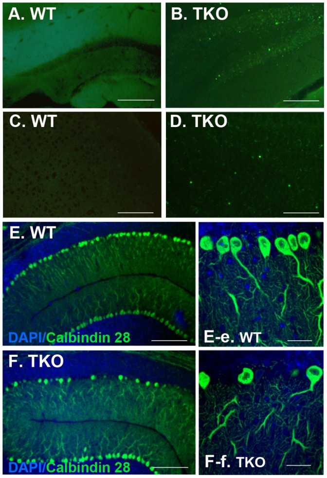
Figure 7. TKO brains show increased apoptosis and cell death.

Mice at 6–8 weeks of age were deeply anesthetized by 2.5% Avertin prior to transcardial perfusion with 1 unit/mL of heparin dissolved in 1×PBS. Brains were harvested, cryosectioned at 20 micron. (A–D) TUNEL labeling showed increased apoptosis in the TKO brains (B and D) as compared to the negative labeling in the WT brains (A and C). (E and F) Purkinje cell loss was shown in the TKO cerebellum (F and F-f) but not in the WT brains (E and E-e). Purkinje cells were immunostained with antibody for calbindin D28K and cell nuclei were stained with DAPI. Fluorescent images were obtained with a confocal microscope (LSM510, Zeiss). Scale Bars in (A–F), 100 µm; and in (E-e and F-f), 50 µm.
