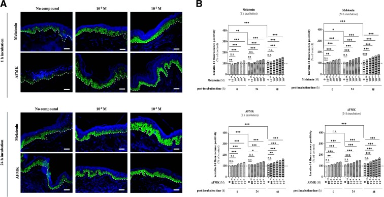
Figure 5.
Effect of melatonin and AFMK on keratin-14 expression. A)Localization of K14 immunoreactivity assessed by immunofluorescence in the human epidermis. Skin samples were incubated over the indicated concentration range with melatonin or AFMK for 1 or 24 h. Following these two incubation times, skin samples were collected immediately (0 h) and 24 or 48 h later, snap-frozen, subjected to cryosections (d=6 μm), and processed for immunolabeling. Detection of the protein was carried out according to the TSA technique as described in Materials and Methods using goat polyclonal anti-K14 IgG (1:400) and secondary biotin-labeled mouse anti-goat IgG (1:400), labeled with FITC (1:50; green). Nuclei were labeled with DAPI solution (blue). Presented images show the K14 expression 48 h after melatonin/AFMK incubation time at specific low (10−8 M) and high (10−3 M) concentrations. Dashed line shows the basement membrane. Scale bars = 50 μm. B) Evaluated data are presented as pooled means ± se of independent experiments showing the effect of melatonin or AFMK at particular concentrations. Values are expressed as a percentage of the control value, i.e., nonpreincubated skin with tested compound at 0 h postincubation. n.s., not significant. *P < 0.05, **P < 0.01, ***P < 0.001.

