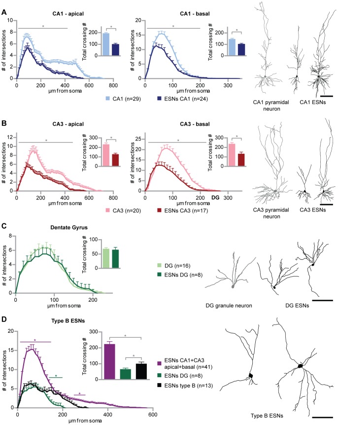
Figure 1. Generation of morphologically different embryonic stem cell-derived neurons (ESN).
(A) ESNs in the CA1 region reproduce the morphology typical of intrinsic CA1 pyramidal cells. Sholl analysis of ESNs in the CA1 region at 21–32 DIV (dark blue) compared to EGFP-f transfected hippocampal neurons at 25 DIV (light blue) for apical and basal dendrites, respectively. (B) CA3 ESNs (dark red) morphologically resemble intrinsic CA3 pyramidal cells (light red) as shown via Sholl analysis and tracing examples. (C) ESNs in the DG (dark green) reproduce the morphology and orientation of intrinsic granule neurons (light green; transfected at 11 and analyzed at 14 DIV) and show the same degree of complexity. (D) Morphologically immature ESNs – type B ESNs – significantly differ from the ESN populations described in (A) to (C) in length and complexity. Numbers of intersections of apical and basal dendrites were totaled in order to compare the different ESN morphologies. Colored significance bars refer to type B ESNs versus ESNs in CA1 and CA3 (purple) and type B ESNs versus ESNs in the DG (green), respectively. For the sake of clarity, significance bars for ESNs in CA1 and CA3 versus ESNs in the DG have been omitted. Student’s t test, *p<0.05. Error bars represent SEM; Scale bars represent 100 µm.

