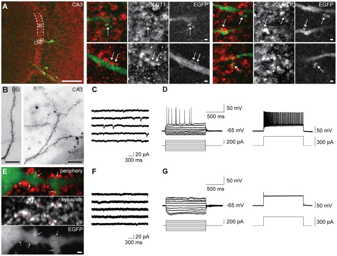
Figure 3. Type A ESNs carry dendritic spines, show EPSCs and fire action potentials.
(A) Co-immunohistochemistry against EGFP and vGLUT1 of type A ESN in the CA3 region of an organotypic hippocampal culture. Detailed images at the right side represent magnification of the four white boxes in the left overview. Arrows point to co-localizations of both markers (sl = stratum lucidum). (B) Type A ESNs in different hippocampal subfields carrying spines. (C) Representative miniature EPSCs of type A ESN in the CA3 region of an OHC. (D) Current-clamp recording from a type A ESN in the CA3 region shows action potentials at distinct levels of depolarization by current pulses (left) and high-frequency firing (right) during a strong depolarization. (E) Type B ESNs are morphologically immature, they do not carry spines. Nevertheless, their dendrites co-localize with the pre-synaptic marker synapsin. ESN found in the periphery of the OHC at 24 DIV. (F) Representative recording showing absence of mEPSCs in type B ESN in the CA3 region. (G) Current-clamp recording showing disability of type B ESN in CA3 region of firing action potentials (left) even during strong depolarization (right). Scale bars in (A) 100 µm (overview) and 1 µm (detailed images), respectively; in (B) 10 µm; (E) 1 µm.

