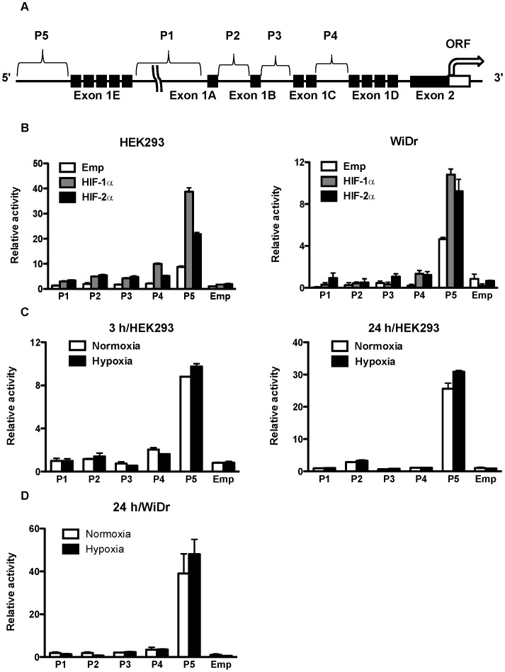
Figure 1. Position of CD133 P5 promoters and their activities after overexpression of HIFs and under hypoxia.
(A) Schematic representation of the position of five CD133 promoters (P1–P5) and exon1s (A–E). (B) Promoter activity of P1, P2, P3, P4 and P5 in human embryonic kidney (HEK) 293 cells (left) and human colon cancer WiDr cells (right) after overexpression of HIF-1α and HIF-2α. (C) Promoter activity of P1, P2, P3, P4, and P5 in HEK293 cells under normoxia and hypoxia for 3 hrs (left) and 24 hrs (right). (D) Promoter activity of P1, P2, P3, P4, and P5 in WiDr cells under normoxia and hypoxia for 24 hrs.

