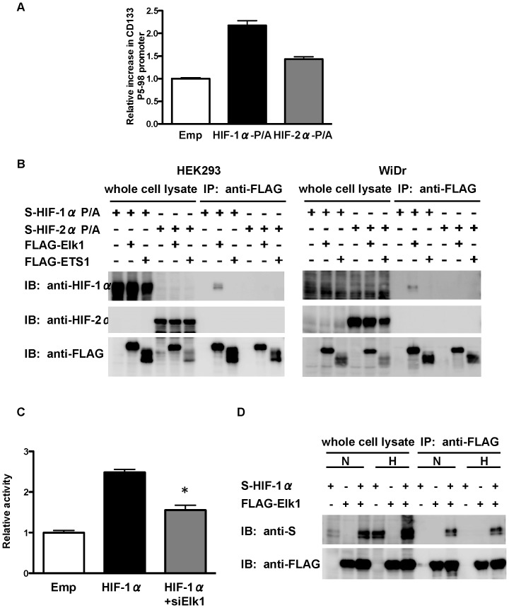
Figure 5. Binding of HIFs to CD133 P5 proximal promoter and ETS-family proteins.
(A) Chromatin immunoprecipitation (ChIP) assay showing the binding of O2-stable HIF-1α and HIF-2α (HIF-1α-P/A and HIF-2α-P/A, respectively) to the CD133 P5 promoter (between −98 bp and +10 bp) in WiDr cells. (B) IP-western blot analysis showing the binding of HIF-1α-P/A and HIF-2α-P/A to ETS1 or Elk1 using human embryonic kidney (HEK) 293 cells (left) and WiDr cells (right). (C) Luciferase activity of P5 −98 bp promoter in HEK293 cells after overexpression of HIF-1α together with the knockdown of Elk1. *P<0.05 vs. HIF-1α overexpression. (D) IP-western blot analysis showing the binding of HIF-1α to Elk1 under normoxia and hypoxia in HEK293 cells.

