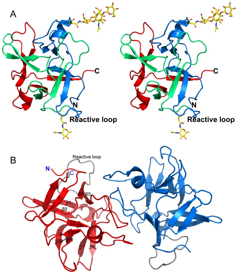
Figure 6. Overall structure of CrataBL.
(A) Stereo view of the three-dimensional structure of a CrataBL monomer (molecule A of crystal form I). Three motifs defining the β-trefoil fold are colored in blue, green, and red. N- and C-termini and the putative reactive loop are labeled. Two glycosylated residues and attached carbohydrates are shown in sticks. The carbohydrate attached to the reactive loop is taken from molecule A in crystal form II. (B) A dimer of CrataBL observed in both crystal forms, with the two molecules colored red and blue, respectively. The reactive loops in both monomers are shown in gray. The secondary structure elements are marked on the molecule shown on the left. Figure prepared with PyMol [69].

