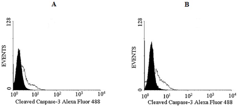
Figure 11. Analysis of caspase-3 activation in prostate cancer cell lines.
DU145 (A) and PC3 (B) (1×10⁵ cells) cell lines were seeded in 6-well plates, following the same protocol for apoptosis with annexin V/FITC and PI staining. Cells treated with CrataBL (40 µM), containing RPMI without FBS were incubated for 48 h at 37°C and 5% (v/v) CO2. The cells were incubated with 10 µL of cleaved caspase 3 Alexa Fluor 488-conjugated antibody for 40 min and analyzed in FACSCalibur flow cytometer. As control, the cells were treated with medium only. The area in black represents the control and in white, cells treated with CrataBL.

